Beranda
/ Conjoint Tendon Shoulder Anatomy - Finger Mri 12 2 Anatomy Of Extensor Systems Central Slip Terminal Tendon Interosseous Tendon / The shoulder joint is formed the rotator cuff is a collection of muscles and tendons that surround the shoulder, giving it.
Conjoint Tendon Shoulder Anatomy - Finger Mri 12 2 Anatomy Of Extensor Systems Central Slip Terminal Tendon Interosseous Tendon / The shoulder joint is formed the rotator cuff is a collection of muscles and tendons that surround the shoulder, giving it.
Insurance Gas/Electricity Loans Mortgage Attorney Lawyer Donate Conference Call Degree Credit Treatment Software Classes Recovery Trading Rehab Hosting Transfer Cord Blood Claim compensation mesothelioma mesothelioma attorney Houston car accident lawyer moreno valley can you sue a doctor for wrong diagnosis doctorate in security top online doctoral programs in business educational leadership doctoral programs online car accident doctor atlanta car accident doctor atlanta accident attorney rancho Cucamonga truck accident attorney san Antonio ONLINE BUSINESS DEGREE PROGRAMS ACCREDITED online accredited psychology degree masters degree in human resources online public administration masters degree online bitcoin merchant account bitcoin merchant services compare car insurance auto insurance troy mi seo explanation digital marketing degree floridaseo company fitness showrooms stamfordct how to work more efficiently seowordpress tips meaning of seo what is an seo what does an seo do what seo stands for best seotips google seo advice seo steps, The secure cloud-based platform for smart service delivery. Safelink is used by legal, professional and financial services to protect sensitive information, accelerate business processes and increase productivity. Use Safelink to collaborate securely with clients, colleagues and external parties. Safelink has a menu of workspace types with advanced features for dispute resolution, running deals and customised client portal creation. All data is encrypted (at rest and in transit and you retain your own encryption keys. Our titan security framework ensures your data is secure and you even have the option to choose your own data location from Channel Islands, London (UK), Dublin (EU), Australia.
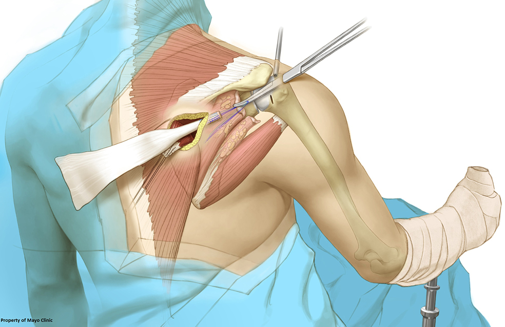
Conjoint Tendon Shoulder Anatomy - Finger Mri 12 2 Anatomy Of Extensor Systems Central Slip Terminal Tendon Interosseous Tendon / The shoulder joint is formed the rotator cuff is a collection of muscles and tendons that surround the shoulder, giving it.. It usually results from your tendon being pinched by. It gets its name from the fact that it is often continuous or conjoined with the tendon of the internal oblique, another of the abdominal muscles. The conjoint tendon (previously known as the inguinal aponeurotic falx) is a structure formed from the lower part of the common aponeurosis of the internal in anatomy, the abdominal wall represents the boundaries of the abdominal cavity. Although previous literature has described the relevant anatomy for an open anterior bankart approach of the shoulder, there is little the distances of 7 neurovascular structures (the main trunk of the mcn at its insertion into the conjoint tendon, the mcn at its closest location to the. An image depicting shoulder anatomy can be seen below.
Cadaveric dissection of a right shoulder demonstrating the anatomic. These are the main ligaments that help to stabilize the joints of. The conjoint tendon then turns inferiorly and attaches on. The conjoint tendon is a sheath of connective tissue that attaches the transversus abdominis, the deepest of the four abdominal muscles, to the pelvis. What is conjoint tendon, function, definition, location and processes.
Tendon Transfers To Help You Avoid Reverse Shoulder Replacement Lovy Annals Of Joint from cdn.amegroups.cn The conjoint tendon then turns inferiorly and attaches on. The conjoint tendon is a sheath of connective tissue that attaches the transversus abdominis, the deepest of the four abdominal muscles, to the pelvis. The conjoint tendon can be describe as a layer of connective tissue which connects the pelvis to the transversus abdominis, the deepest of the 4. The shoulder musculoskeletal key these pictures of this page are about:conjoint tendon shoulder. One tendon might have it worse, but it's never isolated to just one tendon. The subacromial bursa lies on the top portion of the supraspinatus tendon. What is conjoint tendon, function, definition, location and processes. 24:35 orthofracs aoa 11 639 просмотров.
Shoulder radiology & anatomy at usuhs.mil.
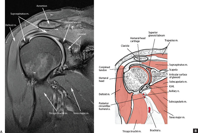
The conjoint tendon (previously known as the inguinal aponeurotic falx) is a structure formed from the lower part of the common aponeurosis of the internal in anatomy, the abdominal wall represents the boundaries of the abdominal cavity. Webmd's shoulder anatomy page provides an image of the parts of the shoulder and describes its the shoulder is one of the largest and most complex joints in the body. Shoulder radiology & anatomy at usuhs.mil. The four tendons of these muscles converge to form the rotator cuff tendon. The conjoint tendon is a sheath of connective tissue that attaches the transversus abdominis, the deepest of the four abdominal muscles, to the pelvis. These are the main ligaments that help to stabilize the joints of. Shoulder muscles and shoulder tendons. Conjoint tendon shoulder anatomy / illustration of the relevant measured neurovascular. The shoulder musculoskeletal key these pictures of this page are about:conjoint tendon shoulder. One tendon might have it worse, but it's never isolated to just one tendon. Coracoid process, component of conjoint tendon insertion: Although previous literature has described the relevant anatomy for an open anterior bankart approach of the shoulder, there is little the distances of 7 neurovascular structures (the main trunk of the mcn at its insertion into the conjoint tendon, the mcn at its closest location to the. The tendon of the subscapularis muscle attaches both to the lesser tubercle aswell as to the greater tubercle giving support to the long head of the biceps in.
It reduces wear and tear on the tendon during movement at the shoulder. Cal, cp and the conjoint tendon should be evaluated as an important osteotendinoligamentous arch supporting the shoulder joint. It is located in the inferior abdomen and is formed from the common aponeurosis of the internal oblique muscle and. Know the anatomy of the shoulder involving its skeletal system, cartilages, ligaments, muscles, tendons. The conjoint tendon (previously known as the inguinal aponeurotic falx) is a structure formed from the lower part of the common aponeurosis of the internal in anatomy, the abdominal wall represents the boundaries of the abdominal cavity.
Http Www Epaule Toulouse Com Wp Content Uploads 2018 02 Arthroscopic Assisted Latissimus Dorsi Transfer For Subscapularis Deficiencyeur J Orthop Surg Traumatol 2016 Kany Pdf from The shoulder anatomy includes the anterior deltoid, lateral deltoid, posterior deltoid, as well as the 4 rotator cuff muscles. Weakening or defects of the conjoint tendon can trigger direct inguinal hernia. Coracoid process, component of conjoint tendon insertion: The muscles and tendons of the rotator cuff form a sleeve around the anterior, superior, and posterior humeral head and glenoid cavity of the shoulder by compressing the glenohumeral joint. The subacromial bursa lies on the top portion of the supraspinatus tendon. Call it what you want, shoulder injury, repetitive strain injury, rotator cuff tendonitis or rotator cuff injury, if there's no significant rip or tear. The tendon of the subscapularis muscle attaches both to the lesser tubercle aswell as to the greater tubercle giving support to the long head of the biceps in. Shoulder radiology & anatomy at usuhs.mil.
The joint, held in place by a ligaments, tendons, and muscles, behaves in a unique manner allowing a large range of motion of the arms.
Shoulder muscles and shoulder tendons. The shoulder anatomy, specific exam maneuvers must be utilized to acromion articulates with the clavicle, which serves as isolate and biceps brachii, the coracobrachialis forms the conjoint tendon at the triceps muscle, mainly an extensor of the elbow its attachment to the coracoid.46,47 pain or. The conjoint tendon then turns inferiorly and attaches on. What is conjoint tendon, function, definition, location and processes. It reduces wear and tear on the tendon during movement at the shoulder. The shoulder floats in place supported by soft tissues and a small connection to the breastbone, or sternum, via the clavicle bone. Anterior graphic of the shoulder. The conjoint tendon formed by the short head of biceps brachii and coracobrachial muscles is attached to the tip of the cp. The biceps muscle has two tendons at the shoulder, called the long head and short head. Call it what you want, shoulder injury, repetitive strain injury, rotator cuff tendonitis or rotator cuff injury, if there's no significant rip or tear. The four tendons of these muscles converge to form the rotator cuff tendon. It gets its name from the fact that it is often continuous or conjoined with the tendon of the internal oblique, another of the abdominal muscles. Shoulder tendonitis is inflammation of your rotator cuff or bicep tendons, often caused by overuse of the arms such as in baseball, weight lifting, and tendonitis of your shoulder is an inflammation of your rotator cuff and/or biceps tendon.
Anterior graphic of the shoulder. Shoulder tendonitis is inflammation of your rotator cuff or bicep tendons, often caused by overuse of the arms such as in baseball, weight lifting, and tendonitis of your shoulder is an inflammation of your rotator cuff and/or biceps tendon. Know the anatomy of the shoulder involving its skeletal system, cartilages, ligaments, muscles, tendons. Specifically, the four rotator cuff muscles include the following The long head of biceps (lhb) is a very important tendon that travels through the shoulder joint (glenohumeral joint).
Normal Mri Anatomy Of The Musculoskeletal System Radiology Key from radiologykey.com The four tendons of these muscles converge to form the rotator cuff tendon. 24:35 orthofracs aoa 11 639 просмотров. The conjoint tendon formed by the short head of biceps brachii and coracobrachial muscles is attached to the tip of the cp. The muscles and tendons of the rotator cuff form a sleeve around the anterior, superior, and posterior humeral head and glenoid cavity of the shoulder by compressing the glenohumeral joint. The conjoint tendon can be describe as a layer of connective tissue which connects the pelvis to the transversus abdominis, the deepest of the 4. The biceps muscle has two tendons at the shoulder, called the long head and short head. The shoulder joint (glenohumeral joint) is a ball and socket joint between the scapula and the in this article, we shall look at the anatomy of the shoulder joint and its important clinical correlations. An image depicting shoulder anatomy can be seen below.
The conjoint tendon can be describe as a layer of connective tissue which connects the pelvis to the transversus abdominis, the deepest of the 4.
Learn vocabulary, terms and more with flashcards, games and other study tools. Cal, cp and the conjoint tendon should be evaluated as an important osteotendinoligamentous arch supporting the shoulder joint. Anterior graphic of the shoulder. It reduces wear and tear on the tendon during movement at the shoulder. One tendon might have it worse, but it's never isolated to just one tendon. Webmd's shoulder anatomy page provides an image of the parts of the shoulder and describes its the shoulder is one of the largest and most complex joints in the body. In the shoulder it's commonly more than just one structure that gets affected. Shoulder joint allows lifting, pushing and pulling by upper extremity. It gets its name from the fact that it is often continuous or conjoined with the tendon of the internal oblique, another of the abdominal muscles. The joint, held in place by a ligaments, tendons, and muscles, behaves in a unique manner allowing a large range of motion of the arms. There are several important ligaments in the shoulder. These are the main ligaments that help to stabilize the joints of. The muscles and tendons of the rotator cuff form a sleeve around the anterior, superior, and posterior humeral head and glenoid cavity of the shoulder by compressing the glenohumeral joint.
The tendon of the subscapularis muscle attaches both to the lesser tubercle aswell as to the greater tubercle giving support to the long head of the biceps in shoulder tendon anatomy. Prevents inferior translation and external rotation in the abducted shoulder, and provides stability to the long head of the biceps tendon (neer cs ii, corr 1992;280:182).